Bringing a new drug to market costs billions of dollars and can take more than a decade. These large investments of money and time contribute to today’s rising healthcare costs as well as significant barriers to bringing new treatments to patients.
One big reason behind these obstacles is the laboratory models that scientists use to develop drugs.
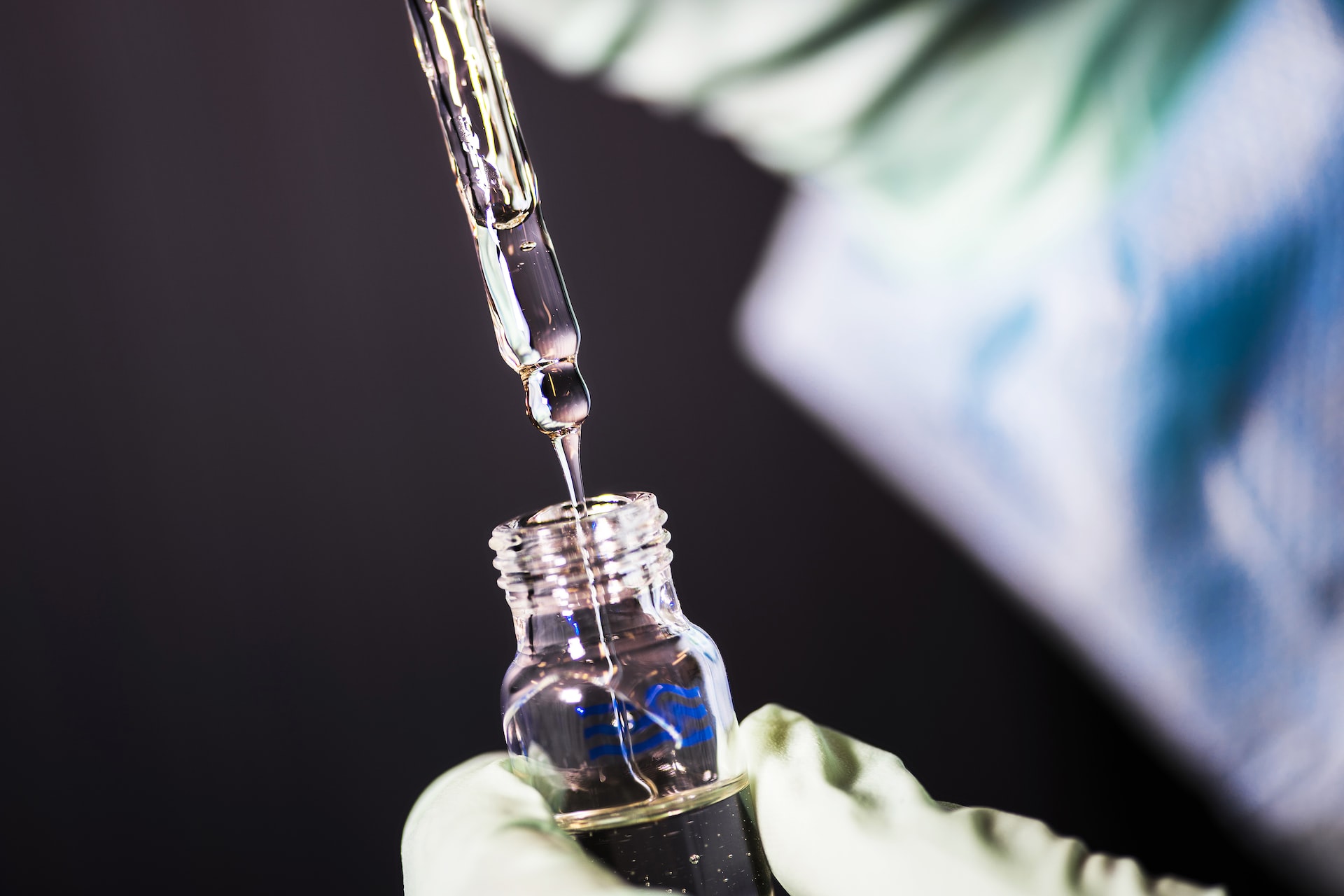
Preclinical studies, i.e., studies that test the efficacy and toxicity of a drug before entering clinical trials in humans, are primarily conducted in cell cultures and animals. Both are limited by their poor ability to mimic the conditions of the human body.
Cell cultures in a Petri dish cannot reproduce all aspects of tissue function, such as the interaction of cells in the body or the dynamics of living organs. And animals are not people—even small genetic differences between species can magnify into large physiological differences.
Less than 8 percent of successful animal studies on cancer treatments make it to human clinical trials. Because animal models often fail to predict drug effects in human clinical trials, these late-stage failures can significantly increase both costs and patient health risks.
To solve this translation problem, researchers have developed a promising model that can better mimic the human body: organs on a chip.

What are intelligent organs?
In the late 1990s, researchers found a way to layer elastic polymers to control and study liquids at the microscopic level. This launched the field of microfluidics, which in biomedical science involves the use of devices that can mimic the dynamic flow of fluids such as blood in the body.
Advances in microfluidics have given researchers a platform to grow cells that work closer to the human body, especially for organs on chips. “Chip” refers to a microfluidic device that encapsulates cells. They are usually made using the same technology as computer chips.
In addition to organs-on-chips that mimic blood flow in the body, these platforms have micro-compartments that allow researchers to integrate multiple types of cells to mimic the types of cells normally present in an organ. Fluid flow connects these multiple cell types, allowing researchers to study how they interact with each other.
This method can overcome the limitations of both static cell cultures and animal experiments in several ways. First, the presence of flowing fluid allows the model to mimic both what a cell experiences in the body, such as how it receives nutrients and removes waste, as well as how a drug moves through the blood and interacts with multiple cell types. The ability to control fluid flow also allows researchers to fine-tune the optimal dose of a particular drug.
For example, a lung-on-a-chip model can combine both the mechanical and physical properties of a living human lung. It can mimic lung expansion and contraction or inhalation and exhalation and simulate the interface between the lungs and air. The ability to reproduce these characteristics allows researchers to better study lung failure under the influence of various factors.

Organ-on-a-chip at scale
Although organs-on-a-chip are pushing the boundaries of early-stage drug research, the technology has not been widely integrated into drug development pipelines.
The main obstacle to the widespread adoption of such chips is their high complexity and low practicality.
Current organ-on-a-chip designs are difficult for the average scientist to use. Moreover, because most models are disposable and allow only one input, limiting what researchers can study in a given time, their implementation is both expensive and time- and labor-intensive. The large investment required to use these models can dampen enthusiasm for their adoption. After all, researchers often use the least complex models in preclinical studies to reduce time and cost.
Lowering the technical bar for the manufacture and use of microchips is critical for the entire research community to take full advantage of their benefits. But this does not necessarily require simplifying the models. For example, my lab has designed a variety of standardized and modular “plug and play” fabrics that allow researchers to easily assemble parts ready for experiments.
The advent of 3D printing has also greatly facilitated the development of organs on chips, allowing researchers to fabricate whole tissues and organ models directly on chips. 3D printing is ideal for rapid prototyping and design sharing between users, and it also facilitates the mass production of standard materials.
Organs on chips can enable breakthroughs in drug development and allow scientists to better understand how organs function in health and disease. Improving the accessibility of this technology could help move the model out of laboratory development and allow it to make its mark in the biomedical industry.
Source: The conversation
Get more news and insights about Global Biotechnology Industry here